Diagnosis & Assessment
It's important to pay attention to changes in your skin, as early detection is a huge factor in the successful treatment of BCC. As BCC does not metastasize, signs and symptoms are limited to the formation of lesions on the surface of the skin. The most common places to find BCC are: the head and neck, the torso and back, and less commonly, arms and legs. BCC can also be reported in unusual sites such as the armpits, breasts, perianal area, genitalia, palms and soles. BCC may present as a pearly white papule (bump) with visible telangiectasias (spider veins), nonhealing scabs, crusts, nonhealing wounds, nodules, plaques, and tumors with rolled borders (Schwartz, 2018).
Assessment typically follows these steps, and is often done by a nurse:
There are various assessment methods used to diagnose BCC. They include..
ACBDEs
Ugly Duckling Sign
EFG Rule
Blue-Black Rule
Assessment typically follows these steps, and is often done by a nurse:
- A thorough history of medical conditions and exposure to sun and UV radiation, since UV radiation is a major risk factor.
- A thorough integumentary (skin) assessment to look for signs of abnormal skin growths. The “stretch test” may be performed to confirm the pearly nature of the lesion by stretching the skin under a light source (Schwartz, 2018).
- If a lesion is found, a shave or punch biopsy is performed with subsequent analysis by a laboratory to confirm whether or not it is cancerous (Schwartz, 2018).
There are various assessment methods used to diagnose BCC. They include..
ACBDEs
- Asymmetry: halves unequal/irregular
- Border: irregular/poorly defined
- Colour: varied with different shades (i.e. tan, brown, black, white, red, blue)
- Diameter: >6 cm (can be smaller)
- Evolving: changing in shape, size, and colour
Ugly Duckling Sign
- Any mole or lesion that stands out from the "flock"
EFG Rule
- Elevated: grown outwards/upwards to form areas of hard lumps
- Firm on palpation
- Growing progressively for a month
Blue-Black Rule
- Recognize any darkly pigmented lesions
- Via dermascope - compare mole or lesion's pattern to 9+ typically benign patterns
- Beauty? Seemingly benign nevus
- Beast? Globules & dots, streaks, eccentric blotches, blue-white veil, negative pigmented network, regression structures, abnormal vascular formations
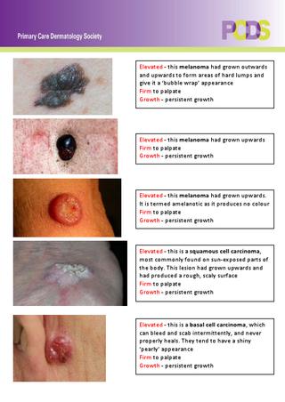
EFG Rule Examples. Primary Care Dermatology Society. (2019)
|
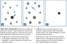
Ugly Duckling Sign. Jensen, J., & Elewski, B. (2015)
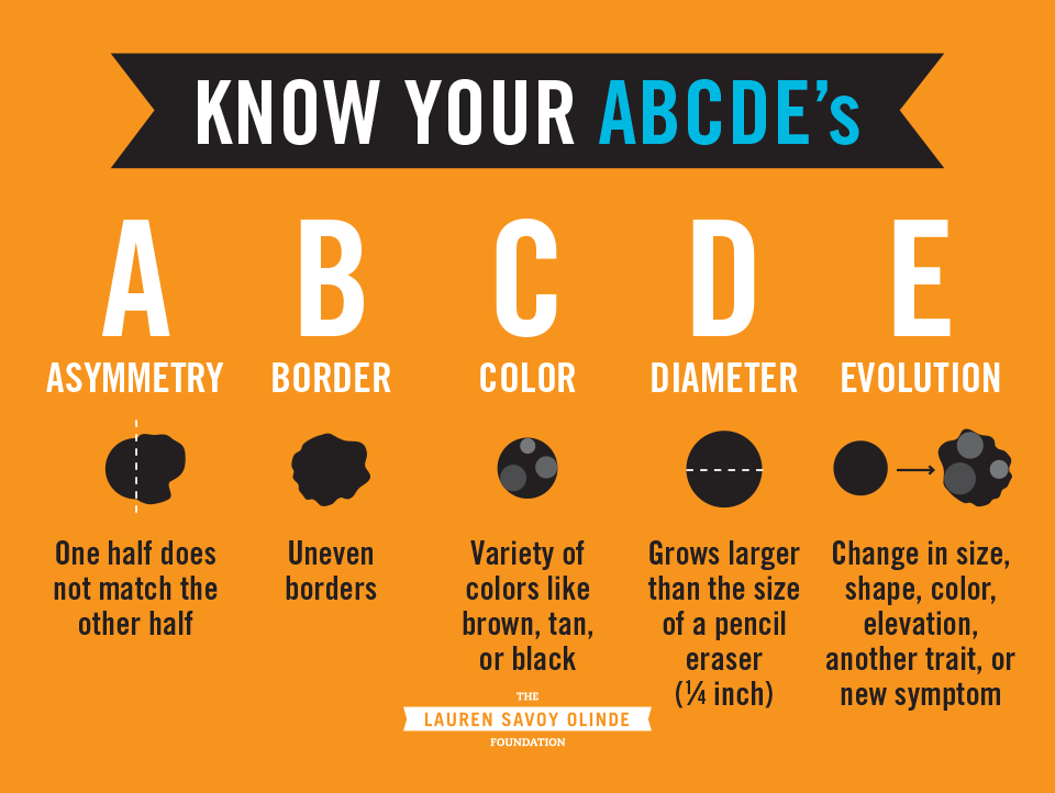
ABCDEs. Hope Health Family Practice. (2019)
|
